Phone: (patient scheduling office): 905-472-7020 When you arrive at Markham Stouffville Hospital: Register at the registration desk in the main lobby. The registration desk clerk will direct you to your procedure.
Uxbridge Hospital:
Phone: 905-852-9771 ext. 5249 When you arrive at Uxbridge Hospital: Register at the central registration desk on the main floor. The registration desk clerk will direct you from there.
Notice
If you’re unable to stand, or need additional support during your appointment, please book your appointment from Monday – Friday, 8:30 a.m. – 3 p.m. so that we can better assist you.
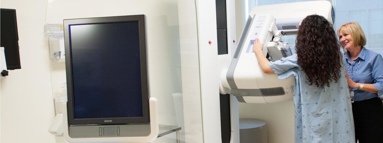
Mammography uses a low-dose X-ray system to examine breasts. A mammogram helps in the early detection and diagnosis of breast diseases in women. Oak Valley Health offers the latest in digital mammography equipment. We use a specially-designed digital camera and a computer to produce an image that is displayed on a high-resolution computer monitor.
Markham Stouffville Hospital also has the latest technology – breast tomosynthesis. During the tomosynthesis scan, multiple low-dose images of the breast are acquired at different angles. These images are then used to produce a series of one millimeter-thick slices that can be viewed as a 3D reconstruction of the breast. Tomosynthesis is used for some women – those with previous breast cancer, with new breast concerns, women who are called back for extra imaging, and those who have a high breast density according to the Ontario Breast Screening Program (OBSP).
Your mammogram imaging will be done by a friendly and professional female registered medical radiation technologist. Your procedure will be reported by a radiologist who specializes in interpreting the results of medical imaging exams.
Diagnostic mammography is used to evaluate a patient with abnormal clinical findings—such as a breast lump or lumps—that have been found by the woman or their doctor.
To have a mammogram, your doctor needs to fill out a referral form.
Screening mammography plays an important part in early detection of breast cancers. It is the best screening test for most women. Women aged 40 to 74 are encouraged to have a mammogram every two years to detect changes in breast tissue that are too small to feel or see.
Regular screening mammograms can find cancer when it is small, which means there is better chance of treating the cancer successfully, it is less likely to spread, and there may be more treatment options. Screening mammograms could save your life.
What is the Ontario Breast Screening Program?
Oak Valley Health is an Ontario Breast Screening Program (OBSP) partners. OBSP is an organized breast cancer screening program that is funded by the Ministry of Health and Long Term Care and managed by Cancer Care Ontario. The OBSP offers important advantages for women and their primary care providers, including scheduling of all screening appointments, sending recall and result letters to women, and arranging follow-up services for women with results that show they need more tests.
The OBSP Program does not require a requisition from your doctor.
Women who are screened for breast cancer within an organized screening program like the OBSP further benefit by participating in a program that undergoes ongoing quality assurance, program monitoring, and evaluation to ensure that its clients receive high-quality screening. In addition, all OBSP sites are accredited with the Canadian Association of Radiologists Mammography Accreditation Program.
Oak Valley Health is accredited with CAR (Canadian Association of Radiologists).
If possible, do not schedule your mammogram for the week before your period if your breasts are usually tender during this time. The best time for a mammogram is one week after your period.
Do not wear deodorant, talcum powder, or lotion under your arms or on your breasts on the day of the exam. These can appear on the mammogram as calcium spots.
If you have previous mammograms that were done at a separate facility, please bring them to your next appointment at Oak Valley Health for the radiologist to view.
Please wear a two-piece outfit on the day of your exam.
During your mammogram, a specially qualified female medical radiation technologist will position your breast in the mammography unit. Your breast will be placed on a special platform and compressed with a paddle (often made of clear Plexiglas or other plastic). The technologist will slowly compress your breast as needed. Our technologist understands that some patients may find this compression slightly uncomfortable, and for this reason we take the utmost care and effort to be as gentle as possible during your mammogram.
Breast compression is necessary to:
Even out the breast thickness so that all of the tissue can be visualized.
Spread out the tissue so that small abnormalities are less likely to be obscured by overlying breast tissue.
Allow the use of a lower X-ray dose since a thinner amount of breast tissue is being imaged.
Hold the breast still in order to minimize blurring of the image caused by motion.
Reduce X-ray scatter to increase sharpness of the pictureYou will be asked to change positions between images. The routine views are a top-to-bottom view and an angled side view. The process will be repeated for the other breast.
A radiologist will analyze and compare your images with previous mammograms. A signed report will be sent to your primary care or referring physician, who will discuss the results with you.
If you are part of the OBSP, the results of your mammogram will be sent to you as well as your family physician within 10 days of your appointment.
When should women have breast screening? The OBSP recommends that women aged 50 to 74 years have a screening mammogram every two years. Women aged 30 to 69 years who have been identified as being at high risk for breast cancer should have a screening mammogram and breast MRI every year. Only those meeting certain criteria are eligible for high-risk screening. To find out more:Download OBSP requisition form.
What is breast density? Breast density refers to the appearance of the fibrous, glandular and fatty tissues on a mammogram. It cannot be determined by the size, firmness, or feeling of the breasts. Density usually decreases gradually after menopause.
If I have dense breasts, do I still need a mammogram? A mammogram is the only medical imaging screening test proven to reduce breast cancer deaths. Many cancers are seen on mammograms even if you have dense breast tissue.
Are there any tests that are better than a mammogram? Studies have shown that ultrasound and magnetic resonance imaging (MRI), combined with mammograms, can aid in diagnosis of breast concerns. However, both MRI and ultrasound show more findings that are not cancer, which can result in added testing, stress, and unnecessary biopsies. Mammography remains to be the gold standard for breast cancer screening for most women.